[big] [huge]
![[big]](screen/01.jpeg)
![[huge]](orig/01.jpeg)
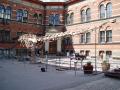
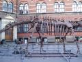
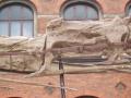
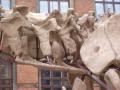
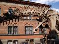
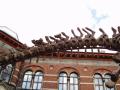
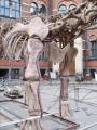
Entire skeletal mount (right anterolateral). It really is as ridiculous as it looks in this photo. Mamenchisaurus is one crazy animal.

[big] [huge]
![[big]](screen/33.jpeg)
![[huge]](orig/33.jpeg)
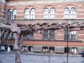
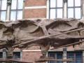
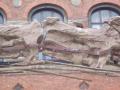
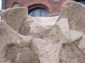
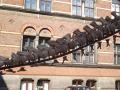
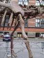
Middle section of neck (anteroventrolateral). This shot didn't quite come off, but was supposed to show how the majority of the cervical vertebrae are twisted clockwise (as you look from the front).
[big] [huge]
![[big]](screen/15.jpeg)
![[huge]](orig/15.jpeg)
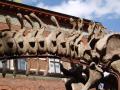
Complete neck (right lateral), consisting of nineteen cervical vertebrae.
[big] [huge]
![[big]](screen/43.jpeg)
![[huge]](orig/43.jpeg)
Anterior half of neck (right lateral), showing C1-12.
[big] [huge]
![[big]](screen/42.jpeg)
![[huge]](orig/42.jpeg)
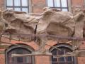
Posterior half of neck (right lateral), showing C13-19 and D1-6.
[big] [huge]
![[big]](screen/40.jpeg)
![[huge]](orig/40.jpeg)
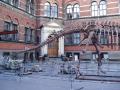
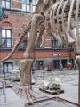
Torso and limbs (right lateral)
[big] [huge]
![[big]](screen/41.jpeg)
![[huge]](orig/41.jpeg)
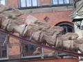
Tail (right lateral). About 47 caudal vertabrae seem to be included, although the resolution of the photograph does not allow an accurate count to made in the distal region. I believe the speciment that this was cast from may have been incomplete in the caudal region.
[big] [huge]
![[big]](screen/05.jpeg)
![[huge]](orig/05.jpeg)
Dorsal column (right lateral). This is a comedy shot, showing how the ribs are mounted to articulate directly with the dorsal central, leaving the diapophyses and parapophyses floating merrily above them. Actually, this isn't such a bad way of mounting a skeleten, since it gives you a better view of the dorsal lamination.
[big] [huge]
![[big]](screen/07.jpeg)
![[huge]](orig/07.jpeg)
Neck and first four cervicals. C4 is broken in half. The head is a Diplodocus cast, quite wrong for Mamenchisaurus, which should have a much more Camarasaurus-like skull.
[big] [huge]
![[big]](screen/08.jpeg)
![[huge]](orig/08.jpeg)
C4-6 and C7a. The rusted metal pole is underneath the break in C6.
[big] [huge]
![[big]](screen/09.jpeg)
![[huge]](orig/09.jpeg)
C7a (posterior part) to C9p.
[big] [huge]
![[big]](screen/10.jpeg)
![[huge]](orig/10.jpeg)
C10a to C11 and anterior part of C12a.
[big] [huge]
![[big]](screen/11.jpeg)
![[huge]](orig/11.jpeg)
C13a (posterior part) to 15a (anterior part).
[big] [huge]
![[big]](screen/12.jpeg)
![[huge]](orig/12.jpeg)
C15a (posteriormost part) to 17 (anterior part)
[big] [huge]
![[big]](screen/13.jpeg)
![[huge]](orig/13.jpeg)
C17-19
[big] [huge]
![[big]](screen/14.jpeg)
![[huge]](orig/14.jpeg)
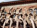
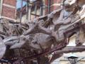
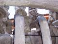
C18-19 and D1-2, massive diapophyses clearly visible. D3 is largely hidden behind the scapula, but its tip is just visible. Comparisons with other photos indicate that its spine slopes backwards, so that there is nothing missing between D2 and the visible dorsal protrusion. The spines of D4 and D5 are visible behind the scapula.
[big] [huge]
![[big]](screen/34.jpeg)
![[huge]](orig/34.jpeg)
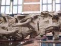
Base of neck (anteroventrolateral), showing cervico-dorsal transition: C16 (posterior part) to C19, D1 to D3.
[big] [huge]
![[big]](screen/06.jpeg)
![[huge]](orig/06.jpeg)
Diplodocus-style skull and C1 to C4a.
[big] [huge]
![[big]](screen/21.jpeg)
![[huge]](orig/21.jpeg)
C5-7 and anterior part of C8
[big] [huge]
![[big]](screen/22.jpeg)
![[huge]](orig/22.jpeg)
C7-9
[big] [huge]
![[big]](screen/23.jpeg)
![[huge]](orig/23.jpeg)
C9-10 and anterior part of C11. From this side, C10 does not appear to be broken, but the fracture is clear from the right.
[big] [huge]
![[big]](screen/24.jpeg)
![[huge]](orig/24.jpeg)
C10p-12a
[big] [huge]
![[big]](screen/25.jpeg)
![[huge]](orig/25.jpeg)
C11 (posteriormost part), C12 and C13a.

[big] [huge]
![[big]](screen/26.jpeg)
![[huge]](orig/26.jpeg)
C12 (posteriormost part), C13 and C14a. C13 has a diagonal crack running posteroventrally through its neural spine.
[big] [huge]
![[big]](screen/28.jpeg)
![[huge]](orig/28.jpeg)
C15 (posterior part), all of C16 and the anteriormost part of C17.
[big] [huge]
![[big]](screen/29.jpeg)
![[huge]](orig/29.jpeg)
C17-19
[big] [huge]
![[big]](screen/30.jpeg)
![[huge]](orig/30.jpeg)
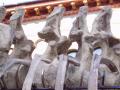
C18-19 and D1; neural arch of D2 visible above the coracoid, and that of D3 above the scapula.
[big] [huge]
![[big]](screen/31.jpeg)
![[huge]](orig/31.jpeg)
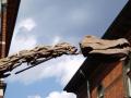
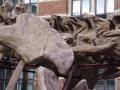
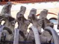
Detail of massive lateral processes on the left side of C18 and C19. Evidently each of these includes a pre- and postzygapophysis, which can be seen in articulation, and a dorsal process. Above that is what may be the left metapophysis of a bifid neural spine, rotated slightly towards the camera so it appears to project more laterally than it should. This strange morphology is not evident at all from in right lateral view, just conventional-looking (though highly laminated) diapophyses and parapophyses, so I may well be completely misinterpreting this.
[big] [huge]
![[big]](screen/35.jpeg)
![[huge]](orig/35.jpeg)
Anterior dorsal column, capturing as much of the vertebrae as possible underneath the scapula. D2-5.
[big] [huge]
![[big]](screen/36.jpeg)
![[huge]](orig/36.jpeg)
Mid-anterior dorsal column, capturing as much of the vertebrae as possible underneath the scapula. D3-5.
[big] [huge]
![[big]](screen/37.jpeg)
![[huge]](orig/37.jpeg)
Mid-posterior dorsal column, D6-9.
[big] [huge]
![[big]](screen/38.jpeg)
![[huge]](orig/38.jpeg)
Posterior dorsal column, D9-12. (Or D12 might be a dorso-sacral S1). It's noticable that the zygapophyses are 4-8cm apart in this region. What does that suggest? The centra seem to fit pretty snugly, so perhaps huge cartilage caps on the zygs?
[big] [huge]
![[big]](screen/39.jpeg)
![[huge]](orig/39.jpeg)
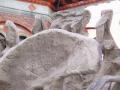
There's very little detail to this shot, as the ilium obscures the view of the sacral vertebrae. D12 is mostly visible; the spines of S1-3 are tightly fused, and S4 is stands alone behind them. Also visible is the spine of Ca1.
[big] [huge]
![[big]](screen/02.jpeg)
![[huge]](orig/02.jpeg)
Proximal half of tail (right posterolateral), showing Ca1-14 and part of Ca15. The chevrons in this region at Y-shaped in anterior view, and simple in lateral view, except for a hint of an anteriorly-directed process on chevron 12/13, which may misplaced. (The spine behind the three fused sacral spines represents the posterior part of the sacrum, S4; the next vertebra behind that is Ca1, so that Ca3-4 appear to ``share'' a neural spine.)
[big] [huge]
![[big]](screen/03.jpeg)
![[huge]](orig/03.jpeg)
Proximal part of tail (right lateral), showing caudals 1-8 and the anterior part of Ca9. It's necessary to count the centra rather than the spines, as the preservation of the latter is poor: e.g. the spines from Ca4-7 are attached to the centra of Ca5-8.
[big] [huge]
![[big]](screen/20.jpeg)
![[huge]](orig/20.jpeg)
Proximal to medial part of tail (right lateral), showing caudals 1-17 and part of Ca18. Chevrons 16/17 and 17/18 are visible forked in lateral view; 15/16 does not appear to be, and shows less anteroposterior flaring at its distal end than does 14/15. As 12/13 shows distinct signs of distal forking, this must be written off either as a preservational artifact or as incorrect mounting.
[big] [huge]
![[big]](screen/04.jpeg)
![[huge]](orig/04.jpeg)
Median third of tail (right lateral), showing the spine of Ca14, all of caudals 15-26 and most of Ca27. The chevrons show a bizarre selection of forked shapes, again suggesting that may have been mounted in the wrong order. Note that the spine of Ca20 is attached to the fused pair Ca21-22.
[big] [huge]
![[big]](screen/32.jpeg)
![[huge]](orig/32.jpeg)
Median caudals Ca10-15 (right lateral), showing poor preservation of neural spines.
[big] [huge]
![[big]](screen/16.jpeg)
![[huge]](orig/16.jpeg)
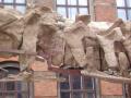
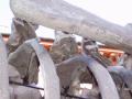
Forelimbs and pectoral girdle (right anterolateral)
[big] [huge]
![[big]](screen/17.jpeg)
![[huge]](orig/17.jpeg)
Forelimbs and pectoral girdle (right lateral)
[big] [huge]
![[big]](screen/18.jpeg)
![[huge]](orig/18.jpeg)
Right hindlimb (right anterolateral)

[big] [huge]
![[big]](screen/19.jpeg)
![[huge]](orig/19.jpeg)
Hindlimbs (right posterolateral)